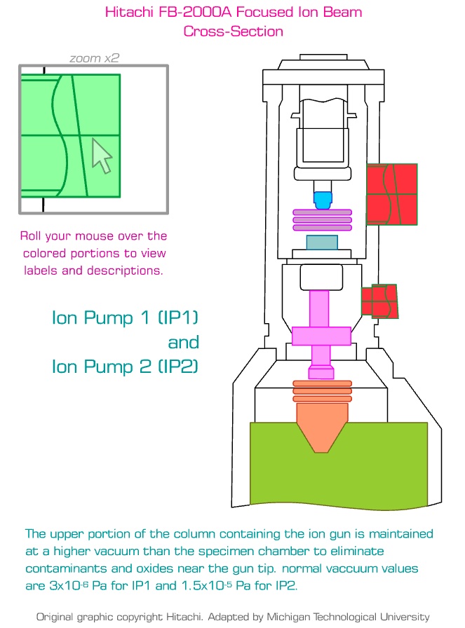
Ion Pump 1 (IP1) and Ion Pump 2 (IP2)
The upper portion of the column containing the ion gun is maintained at a higher vacuum than the specimen chamber to eliminate contaminants and oxides near the gun tip. Normal vacuum values are 3x10⁻⁶ Pa for IP1 and 1.5x10⁻⁵ for IP2.
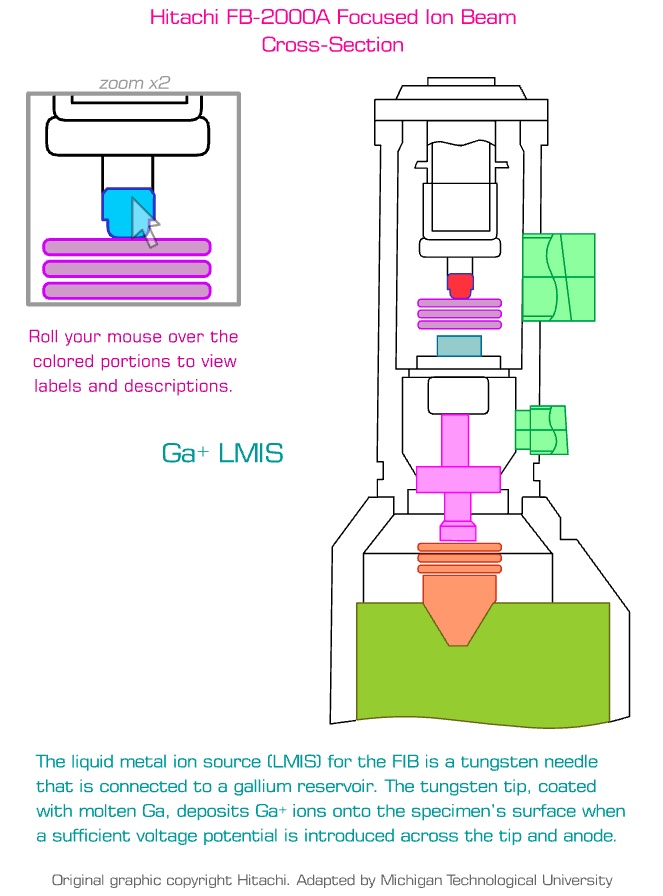
Ga+ Liquid Metal Ion Source (LMIS)
The LMIS for the FIB is a tungsten needle that is connected to a gallium reservoir. The tungsten tip, coated with molten Ga, deposits Ga+ ions onto the specimen's surface when a sufficient voltage potential is introduced across the tip and anode.
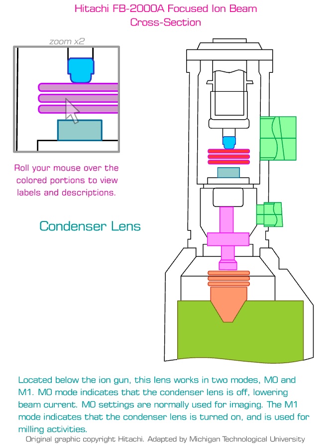
Condenser Lens
Located below the ion gun, this lens works in two modes, M0 and M1. M0 mode indicates that the condenser lens is off, lowering beam current. M0 settings are normally used for imaging. The M1 mode indicates that the condenser lens is turned on and is used for milling activities.
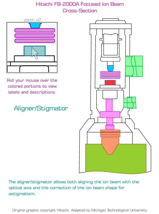
Aligner/Stigmator
The aligner/stigmator allows both aligning the ion beam with the optical axis and the correction of the ion beam shape for astigmatism.
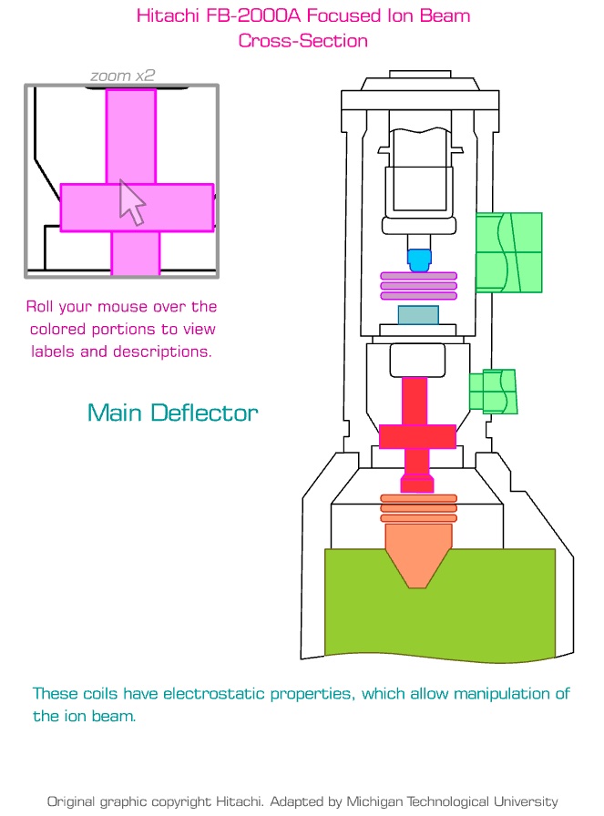
Main Deflector
These coils have electrostatic properties, which allow manipulation of the ion beam.
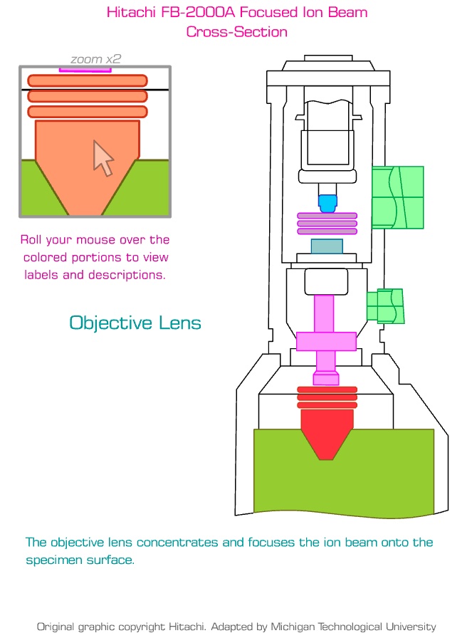
Objective Lens
The objective lens concentrates and focuses the ion beam onto the specimen surface.

Specimen Chamber
The specimen chamber is the enclosed area under vacuum and beneath the ion column where the specimen is located. The normal vacuum value is 5x10⁻² Pa. This chamber encloses the specimen stage.
FIB Internal Components Tour (24 seconds, no audio)
This video provides a virtual, top-to-bottom tour of the internal components within the vacuum column of the Hitachi FB-2000A Focused Ion Beam (FIB) system. The user rolls their mouse over various portions of the system and labels and descriptions are provided.
As the tour moves down the internal ion column, it highlights the following scientific components in sequence:
-
Ion Pump 1 (IP1) and Ion Pump 2 (IP2): Located at the upper portion of the column, these pumps maintain a high vacuum environment (higher than the lower specimen chamber) to eliminate contaminants and oxides near the ion gun tip.
-
Ga+ Liquid Metal Ion Source (LMIS): Positioned at the very top, this is the source of the ion beam. It consists of a tungsten needle connected to a gallium reservoir. When a voltage is applied, the molten gallium coating the tip deposits Ga+ ions to create the beam.
-
Condenser Lens: Moving downward, the beam passes through the condenser lens. This lens dictates the beam's current and operates in two modes: M0 mode (lens off, used for low-current imaging) and M1 mode (lens on, used for high-current milling).
-
Aligner and Stigmator: Just below the condenser lens, this component aligns the focused ion beam with the column's optical axis and corrects the shape of the beam to prevent astigmatism.
-
Main Deflector: This section contains electrostatic coils that precisely manipulate and steer the ion beam's path.
-
Objective Lens: Near the bottom of the column, the objective lens concentrates and focuses the manipulated ion beam directly onto the surface of the specimen.
-
Specimen Chamber: The tour concludes at the bottom in the enclosed specimen chamber. Kept under standard vacuum, this area houses the specimen stage where the focused beam ultimately interacts with the target material for imaging or micromachining.
Original graphic copyright Hitachi. Adapted by Michigan Technological University.