Operation
Ion Source
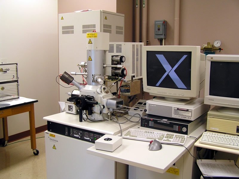
Unlike the electron beam used in scanning and transmission electron microscopy (SEM, TEM), the beam in a focused ion beam system (FIB) is composed of gallium (Ga) ions. The ions forming the structure of the beam are derived from a liquid metal ion source (LMIS). In the Hitachi FB-2000A FIB, the ion source is a tungsten needle that is connected to a gallium reservoir. The accelerating voltage in the Hitachi FB-2000A FIB is adjustable; however, it always operated at 30 kV. Gallium proves to be an exceptional ion source because it not only avoids interactions with tungsten, but it also provides a stable flow of ions at room temperature.
Sputtering
Under the influence of the ion beam, atoms within the specimen surface are ejected due to collision cascades in the target, escaping from the outside layer and into the vacuum. This process is known as sputtering and is carried out in a precise manner in the FIB. In practice, a pattern is constructed in a special computer-aided design (CAD) program. Once an area of the specimen is selected, the ion beam is positioned on the sample based on the CAD diagram.
Imaging
Secondary electrons (SE), emitted from the ion beam-specimen interaction volume, are used for imaging. As the ion beam scans the specimen, the detector simultaneously collects the SEs. Imaging electronics identify the secondary electrons and synchronize that signal with the beam position. Differences in the specimen topography are displayed as varying contrast levels in the image.
The image resolution at the standard 5 mm working distance can reach 5 nm. Non-conductive specimens are sometimes problematic and produce charging effects. Specimen charging occurs when the specimen material cannot dissipate the beam particles, and the resulting image may contain regions of abnormality and/or high contrast. Generally, insulating regions appear dim and conductive regions appear bright.
It is very important to note that the Hitachi FB-2000A FIB is a single beam FIB; therefore, the ion beam is the source of sputtering and secondary electrons used for imaging. This is a drawback to single beam FIBs since, even at very low beam currents, some milling occurs during imaging.
Vacuum
As with an electron microscope, the FIB functions under a vacuum that removes air and contaminants from the ion column. The vacuum environment in a high vacuum instrument precludes the direct examination of living things, oils, or anything that contains liquids. Samples must be dried before use in the system. The high-energy beam interacts with air and hydrocarbons on surfaces in the column and specimen chamber, "pasting" the contamination onto parts in the column. This process eventually degrades the optical qualities of the microscope and images. For this reason, all specimens and specimen holders must be kept clean and should be handled with gloves.
Commonly used vacuum systems consist of an oil-filled roughing pump to produce a vacuum level sufficient to operate the oil diffusion pump (DP) for high vacuum. The roughing pump evacuates the microscope column from atmosphere to low vacuum. The DP then creates the higher vacuum needed for the FIB to operate under standard conditions. Ion (or ion-getter) pumps are used in the gun area of the column. The DP does not remove all gases in the gun/column area. Those molecules are ionized in the ion pump and trapped on a metal surface, creating a pumping action. A sufficient vacuum level must be present before activation of the ion pump. The Hitachi FB-2000A FIB contains three pumping regions. One region is for the ion source and ion column, another is for the detectors and specimen, and yet another is for the specimen exchange chamber. The typical vacuums are 1x10⁻⁸, 1x10⁻⁶, and 1x10⁻⁴ torr, respectively.
Lenses and Detectors
The radius of a gallium ion used to mill specimens in the FIB is 1000 times greater than that of an electron. For this reason, electromagnetic lenses used in electron microscopes would have little effect on the ion beam. The Hitachi FB-2000A FIB uses electrostatic lenses that focus the beam of heavy Ga ions. The condenser lens, which is near the gun, regulates the beam diameter and current. The lower, or objective lens, positions and focuses the beam onto the sample.
As mentioned earlier, secondary electrons are the only imaging signal used in the Hitachi FB-2000A FIB. The FIB uses a variation on the Everhart-Thornley (ET) secondary electron detector. Secondary ions are also produced in the beam-specimen volume and can be used for imaging; however, the Hitachi FB-2000A FIB at Michigan Tech is not equipped to sense them.