
Fluorescent dyes help chemists see the inner workings of disease.
Dysfunctional mitochondria have been linked to neurological and cardiovascular diseases, such as Alzheimer’s and Parkinson’s disease, and even to some types of cancer. To keep these powerhouses working efficiently, cells remove damaged mitochondria. This process, called mitophagy, is like a cell taking out the trash. In diseased cells, the garbage piles up and the cell’s pH changes.
Be Brief Series
Sometimes, you just have to lay eyes on research; a photo is worth a whole essay.
To watch the mitophagy process closely, chemist Haiying Liu collaborated with biologists to make a glowing dye that shines brighter under certain pH conditions that mimic changes caused by disease. The team uses a rhodol-based dye, which is sensitive, permeates cells well and shows low cell toxicity.
“It is important to track the mitophagy process and we need a tool to track it,” Liu said, explaining that mitophagy is not readily visible to the naked eye or even under a microscope.
About the Researchers
Haiying Liu (Chem), Rudy Luck (Chem), Thomas Werner (Bio Sci), Yibin Zhang (research scientist), undergraduate students Logan Miksell, Nick Whisman and Tessa E. Steenvinkel, and graduate students Shuai Xia and Mingxi Fang.
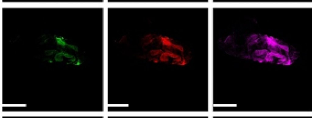
In order to track and visualize the mitophagy process, two different mitochondria- and lysosome-targeting fluorescent probes with different emission wavelengths would be needed to detect the dramatic pH changes during mitophagy: a slightly basic pH of 8.0 in the mitochondria and an acidic pH of 4.5 in the autolysosomes.
The rhodol dyes emit in both visible and the near-infrared regions with self-calibration capability and can effectively track the mitophagy. Liu hopes they will one day work in near-infrared spectra to take advantage of near-infrared imaging such as deep-tissue penetration, the smallest water Raman peak, very little background fluorescence and minimal photo damage to cells and tissues. The end goal: “We hope to make a tool that is easy for industry to prepare and use.”
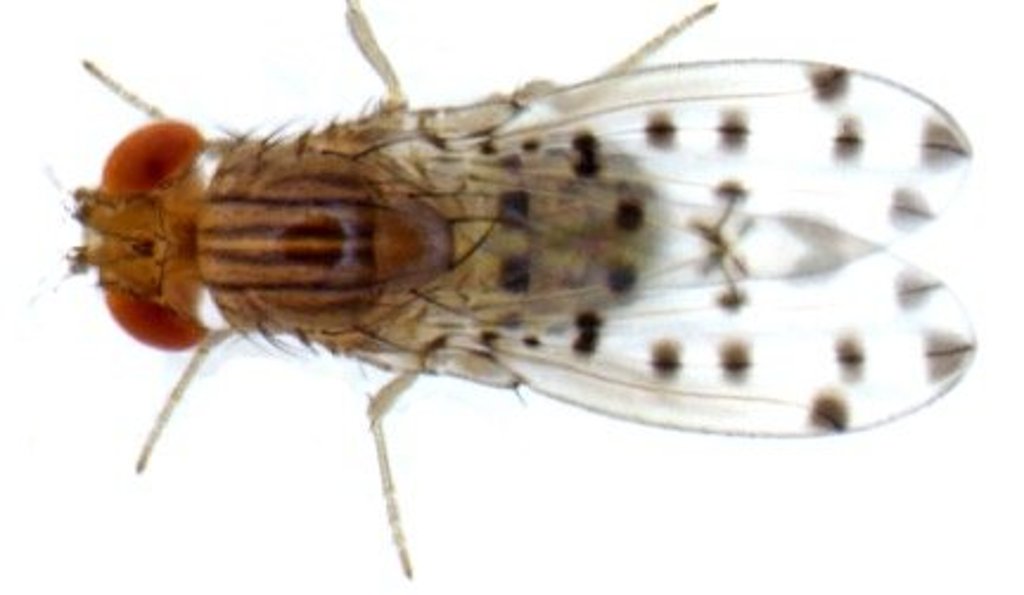
Those potential uses include assessing drug treatments, drug interactions, a better understanding of cellular function and disease monitoring. The team examined how well the dye worked in live cells and fruit fly larvae. The next steps include modification of rhodol dyes to develop water-soluble, near-infrared theranostic prodrugs to precisely target cancer cells, deliver therapeutic drugs to tumors, improve the therapeutic efficacy, and significantly reduce toxicity to normal cells through real-time fluorescence monitoring of drug delivery process.
Michigan Technological University is an R1 public research university founded in 1885 in Houghton, and is home to nearly 7,500 students from more than 60 countries around the world. Consistently ranked among the best universities in the country for return on investment, Michigan's flagship technological university offers more than 120 undergraduate and graduate degree programs in science and technology, engineering, computing, forestry, business, health professions, humanities, mathematics, social sciences, and the arts. The rural campus is situated just miles from Lake Superior in Michigan's Upper Peninsula, offering year-round opportunities for outdoor adventure.

Comments