Biofluids Lab News
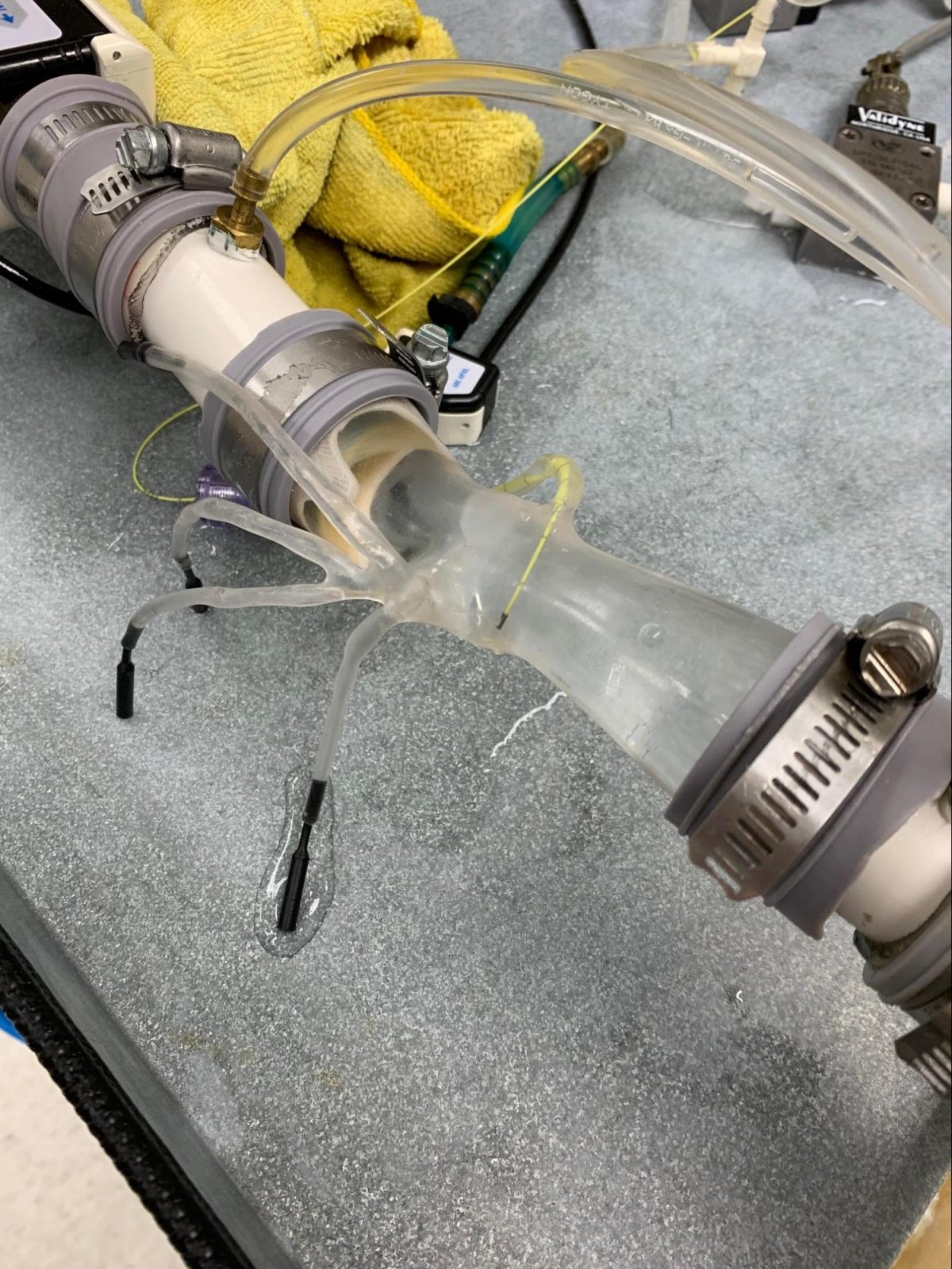
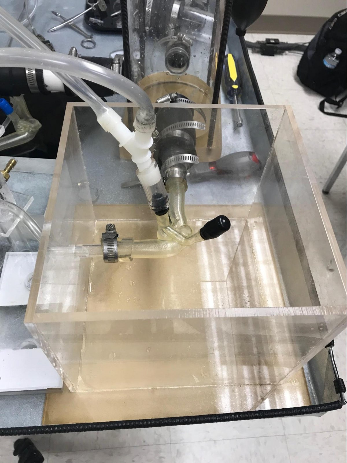
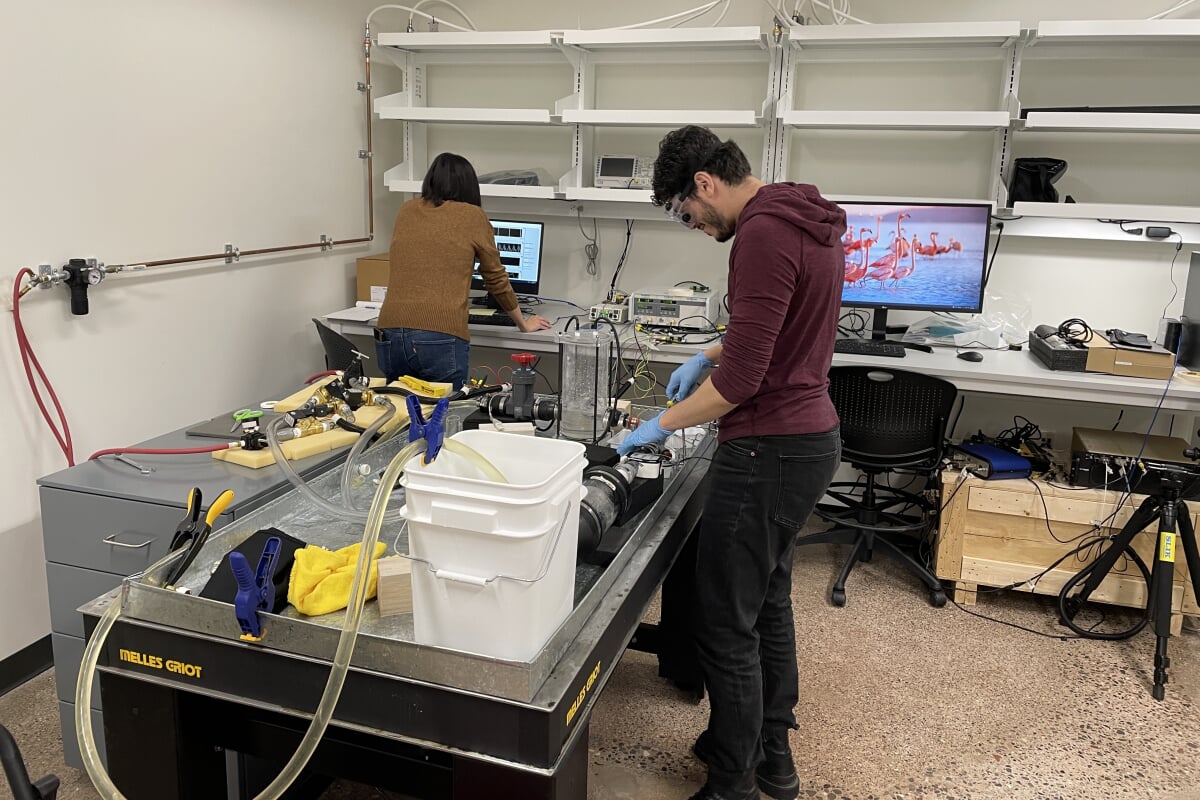
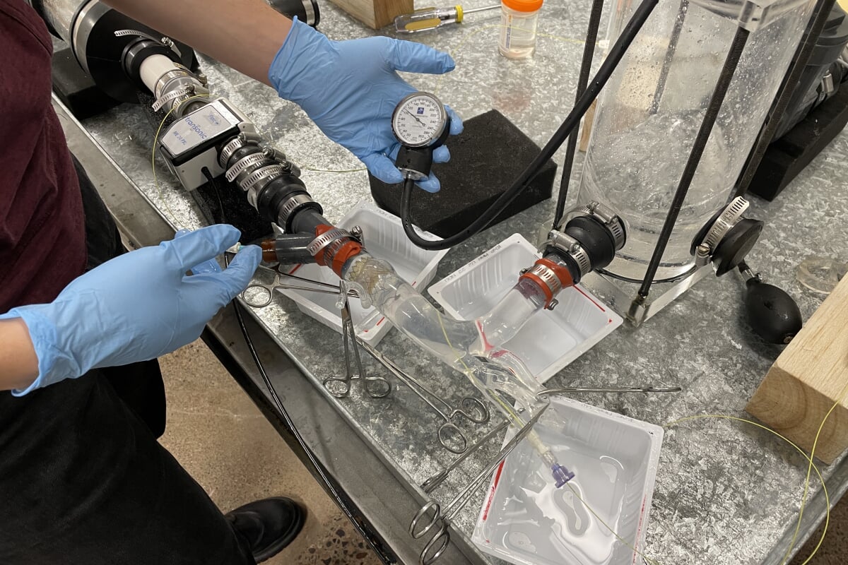
The biofluids lab utilizes fluid and solid mechanics principles, clinical expertise and design and manufacturing to find solutions for cardiovascular flow problems. The lab scope involves both basic and translational studies. The lab utilizes a custom-designed and built pulse duplicator system that emulates that of the cardiovascular system. In this flow simulator setup, hemodynamic assessment of adult and congenital heart defects can take place.
Special equipment used in the lab involves a particle image velocimetry system that allows the characterization of the flow field in vessels and organs.
Location: H-STEM 304E

Hoda Hatoum
- Assistant Professor, Biomedical Engineering
- Affiliated Assistant Professor, Mechanical and Aerospace Engineering
- hhatoum@mtu.edu
- 906-487-2852
- H-STEM 352
Atrial Fibrillation
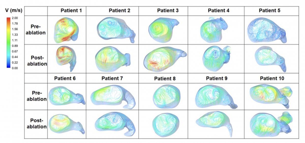
Atrial Fibrillation (AF) is an irregular heart rhythm that is believed to initiate in the atria. An approach to address AF is AF ablation. AF ablation uses heat or cold energy to create scarring around the pulmonary veins to prevent irregular electrical impulses from propagating and creating the arrhythmia. In our lab, we aim to understand the effect of AF and AF ablation approaches on the flow dynamics, to correlate the flow dynamic parameters to clinical outcomes, and to understand the occurrence of stroke as a result of AF.
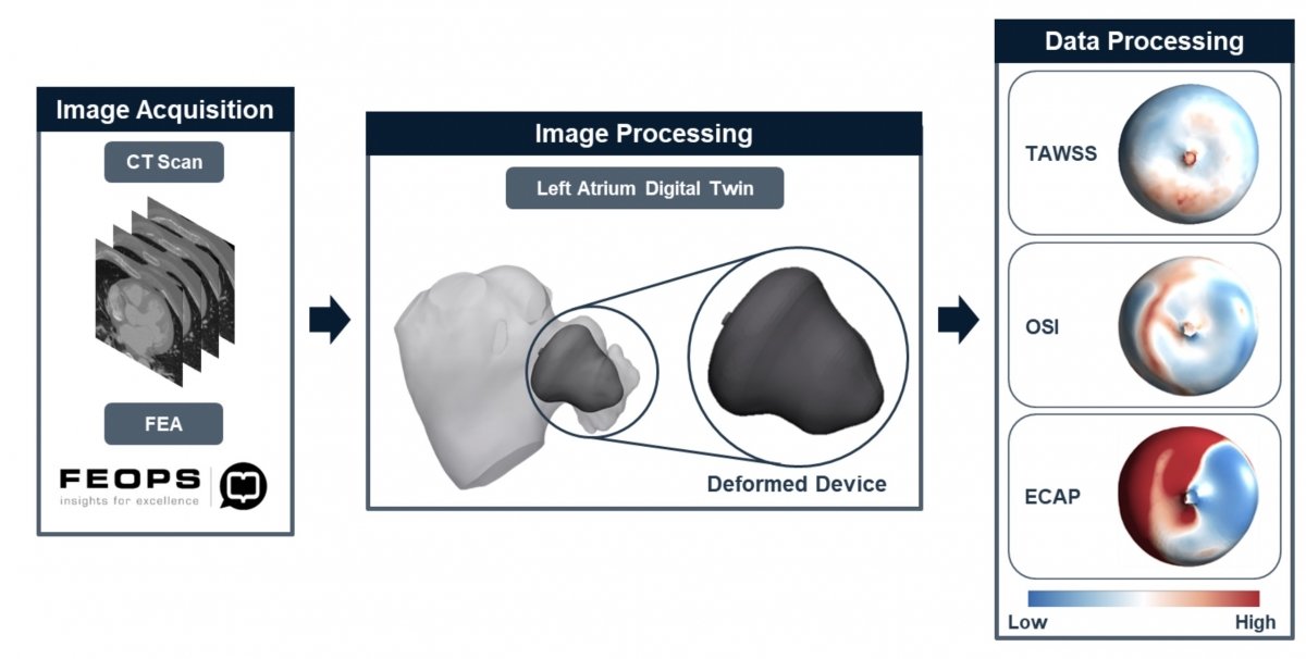
We are also interested in investigating left atrial appendage occlusion (LAAO), specifically the factors that lead to device related thrombosis (DRT).
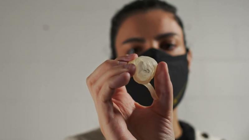
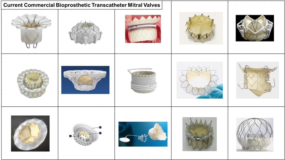
Heart Valve Design
In the biofluids lab, we design heart valve devices. With the rise of minimally invasive surgeries, the clinical field is moving towards transcatheter approaches. Currently, transcatheter heart valves are made of biological materials that are prone to degeneration leading to compromised valve performance and necessitating another valve replacement ultimately. In the biofluids lab, we collaborate with material science experts to utilize novel biomaterials that are biocompatible, durable and suitable for cardiovascular applications. We also design and optimize stents and leaflet geometries.
Heart Valve Performance Assessment
In the biofluids lab, we assess the performance and the flow characteristics of different heart valves in idealized chambers or in patient-specific ones. Using our heart simulator and our particle image velocimetry system, multiple commercially available or in-house made heart valves are tested. Valves are tested for (1) Overall performance and energetics (2) turbulence generated (3) sinus hemodynamics (aortic and pulmonic) and (4) ventricular, atrial, pulmonic and aortic flows.
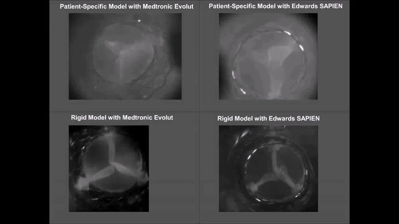
In this video, high-speed imaging was taken of 2 commercial transcatheter aortic valves deployed in an idealized model and in a patient-specific model.
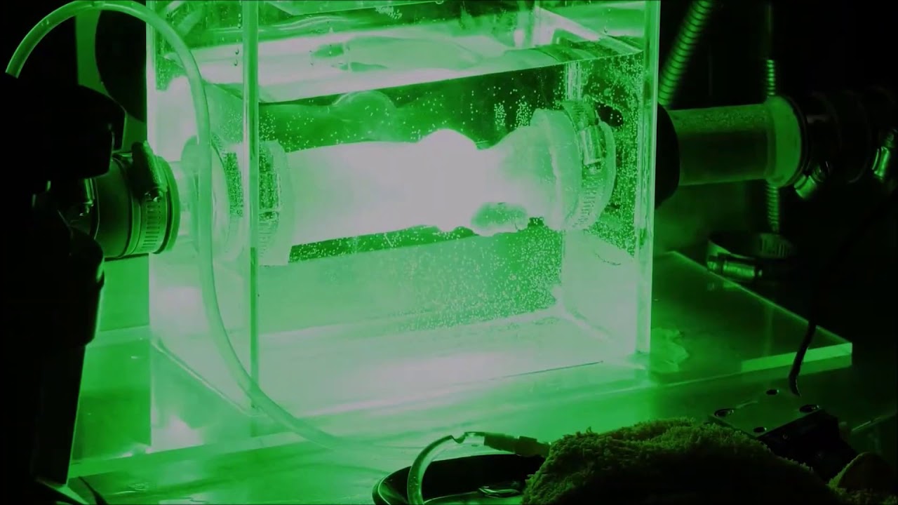
This video shows a particle image velocimetry (PIV) experiment. Using PIV, we shine a laser beam on the region that we are interested in assessing. In our experiments, we utilize a blood analog that is transparent. The fluid is seeded with fluorescent particles that shine upon exposure to the laser beam. Using a specific software, we can calculate the resulting displacement, velocity and any other parameters that we are interested in.
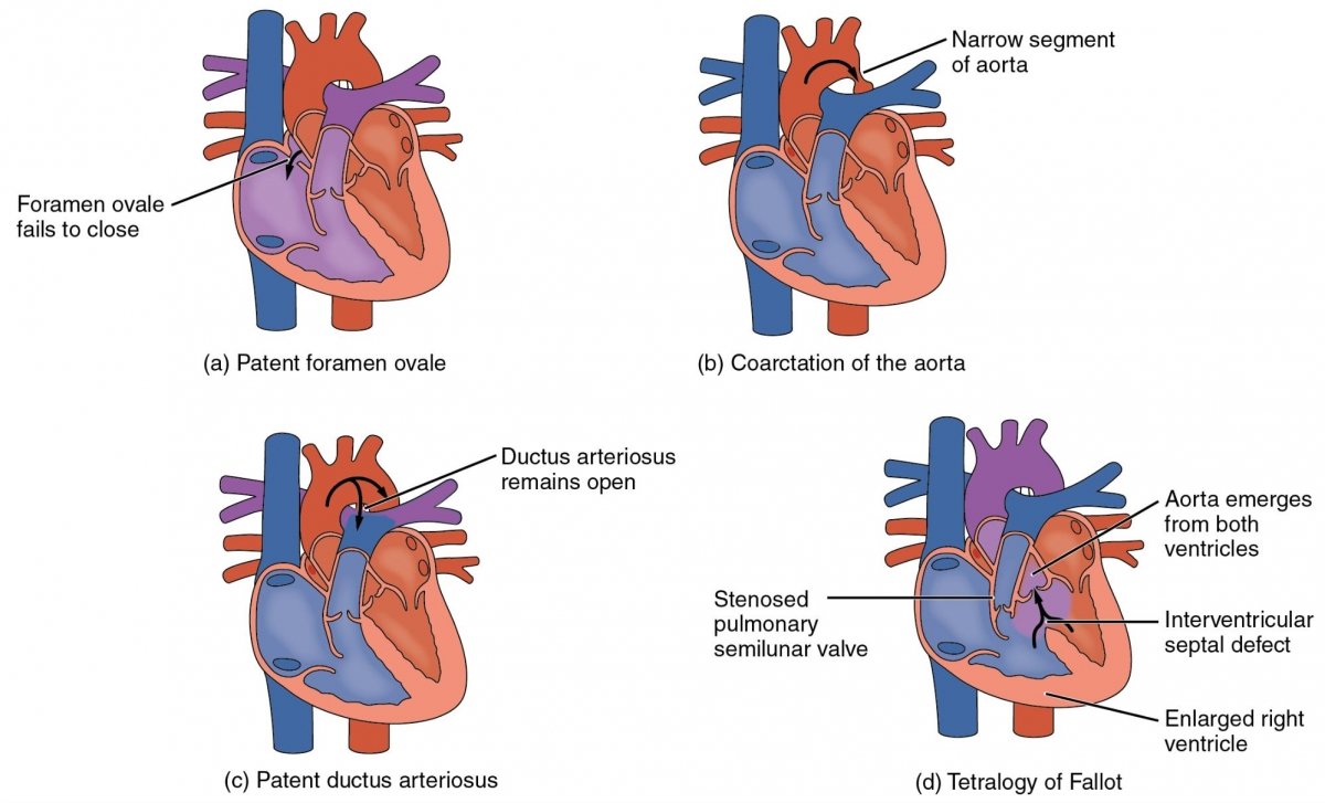
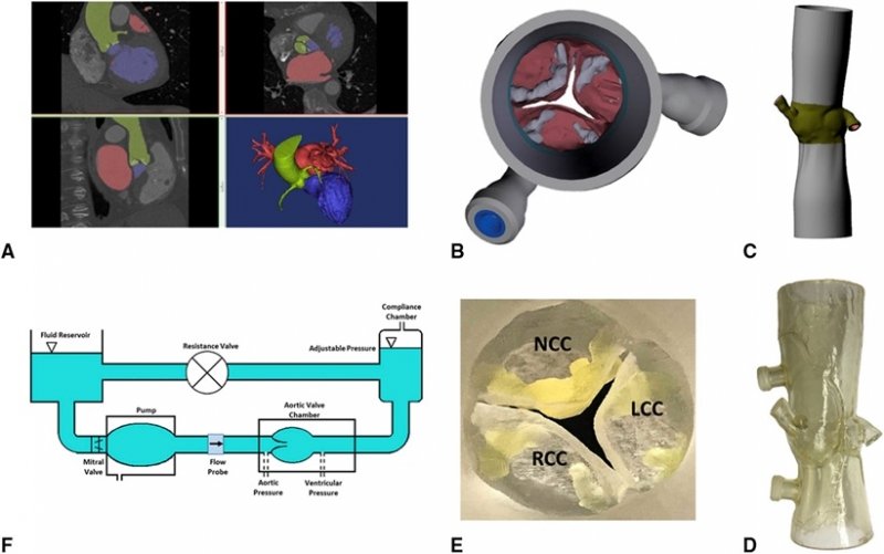
Congenital Heart Defects
In the biofluids lab, we analyze several congenital heart defects and we devise various approaches to help find alternatives for highly-invasive surgeries (Tetralogy of Fallot, pulmonary atresia, double outlet right ventricle, Kawasaki disease, etc.). We collaborate with Nationwide Children’s Hospital to acquire patient data. Using experimental and computational fluid dynamics, we assess different scenarios of design approaches and we develop predictive models to help clinicians with anticipating adverse outcomes.