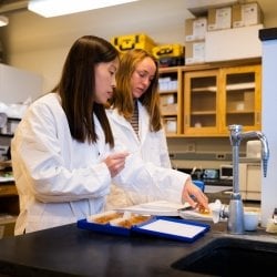
Biofluids Laboratory
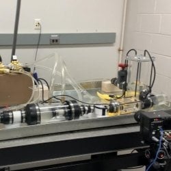
The Biofluids Lab utilizes fluid and solid mechanics principles, clinical expertise, design, and manufacturing to find solutions for cardiovascular flow problems. The lab scope involves both basic and translational studies. The lab utilizes a custom-designed and built pulse duplicator system that emulates that of the cardiovascular system. In this flow simulator setup, hemodynamic assessment of adult and congenital heart defects can take place.
Special equipment used in the lab involves a particle image velocimetry system that allows the characterization of the flow field in vessels and organs.
Location: H-STEM 304E
Contact: Hoda Hatoum
Biomedical Microdevices Lab
Micro- and nano- structures have a significant contribution in the physical world, despite their seemingly insignificant dimensions. The Biomedical µDevices Lab focuses on investigating the microscopic structures that affect physiology and cell biology. Our multidisciplinary research extends to understanding and manipulating the microenvironment to study both cancer metastasis and wound healing.
Location: HSTEM Complex 257
Contact: Smitha Rao
Biomedical Optics Laboratory
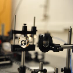
Research in the Biomedical Optics Laboratory is concerned with the way light interacts with human tissue and how this interaction can be used for developing novel ways to image physiological processes and anatomical structures, for developing new laser-based optical diagnostic tools, and even for developing new ways to use light in the treatment of disease. The lab is a fully equipped, modern optics facility that houses vibration-isolated optical benches, numerous types of lasers and other light sources, optoelectronic equipment, optical components, computers for conducting numerical simulations and for collecting and analyzing data, and an area for preparing and studying biological tissues and samples.
Location: Minerals and Materials 114/115
Contact: Sean Kirkpatrick
Brain Stimulation Engineering Lab
Our mission is to engineer novel brain stimulation paradigms through technology development, computational modeling, and experimental studies, with the goal of advancing the therapeutic efficacy and efficiency of deep brain stimulation for the treatments of neurological diseases.
Contact: Chunxiu (Traci) Yu

Bruce Lee Research Group
The Lee Group research is focused on applying biologically-inspired molecular designs with chemistry, polymer engineering, and materials science principles in developing advanced and functional materials for various biomedical applications. Current projects include applying biomimetic structural designs to create tough hydrogels that can potentially function as tissue adhesives or extracellular matrices for tissue repair and regeneration, hydrogel actuators, and smart adhesives.
Contact: Bruce Lee
Mechanobiology Lab
We study mechanobiology, particularly on how adherent cells can sense and respond to mechanical stiffness of the extracellular matrix. To investigate this, we have established experimental and computational frameworks for force measurement and adhesion dynamics quantification. We apply these frameworks, with cutting edge computer vision techniques, on live-cell microscope images to find out the fundamental mechanism underlying mechanosensation in the normal cells and the biomechanical signature in the diseased cells whose signaling has gone awry.
Location: HSTEM Complex 357
Contacts: Sangyoon Han
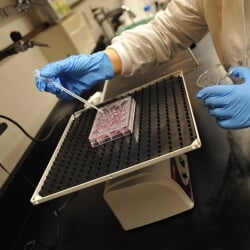
Tissue Engineering and Biomaterials Facility
Our activities include mammalian cell culture, tissue culture, bioreactor design and operation, biomaterial fabrication, and polymer synthesis.
Location: HSTEM Complex 255
Contacts: Bruce P. Lee
Vascular Engineering Laboratory
Laboratory activities are focused on understanding how reduced interstitial flow, following lymphatic injuries, alters lymphatic regeneration and recovery of lymphatic function. The long-term goal of this research is to develop novel biomaterial and tissue engineering strategies for increasing interstitial flow, thereby improving lymphatic regeneration.
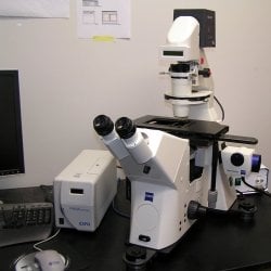
The lab is equipped for small-animal surgeries, tissue cryosectioning, fluorescence and brightfield microscopy, and live lymphatic imaging. Specialized instruments include an Olympus BX51 microscope and a Zeiss Apotome fluorescence microscope—both equipped with a high-resolution color digital camera—two Olympus stereo microscopes, available for small-animal surgeries with affixed DP71 digital color cameras, and a Near Infrared (NIR) imaging system, available for live imaging of lymphatic vessels in both rodents and humans.
Location: HSTEM Complex 357
Contact: Jeremy Goldman