
From energy materials to disease diagnostics, new microscopy techniques can provide more nuanced insight. Researchers first need to understand the effects of radiation on samples.
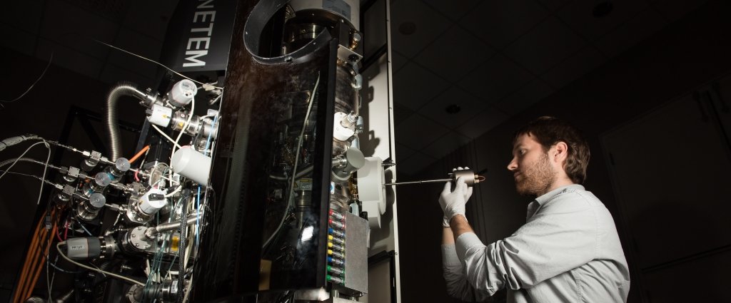
In a new paper published last week in Science Advances (DOI: 10.1126/sciadv.aaq1202), a team of scientists and engineers dug into the mechanisms that degrade sample quality in liquid cell transmission electron microscopy (LC-TEM). They developed an LC-TEM device that uses multiple windows and patterned features to explore the impacts of high-energy electron bombardment on nanoparticles and sensitive biological samples.
The collaborating institutions include the EMSL, the Environmental Molecular Sciences Laboratory, a Department of Energy Office of Science User Facility at the Pacific Northwest National Laboratory (PNNL), University of Illinois Chicago, Florida State University, Washington State University and Michigan Technological University. The study's lead author, Trevor Moser, currently at the PNNL, is a doctoral student at Michigan Tech studying under both Tolou Shokuhfar, an adjunct professor of mechanical engineering at Michigan Tech and an associate professor of bioengineering at the University of Illinois Chicago, and James Evans, a senior scientist at PNNL.
The team explains that transmission electron microscopy (TEM) relies on a high-energy beam of electrons that passes through a sample. Whether the sample is from a battery electrode or bacterial cells, the passing electrons will scatter in a specific way reflecting the sample's atomic structure. In LC-TEM, materials can be examined in a native state allowing dynamic observations, but the samples are liquid or suspended in liquid and have to be tightly sealed to withstand the space-like vacuum of the instrument. There is a balance between ensuring the liquid doesn't evaporate while providing enough viewing space for the electron beam to pass through.
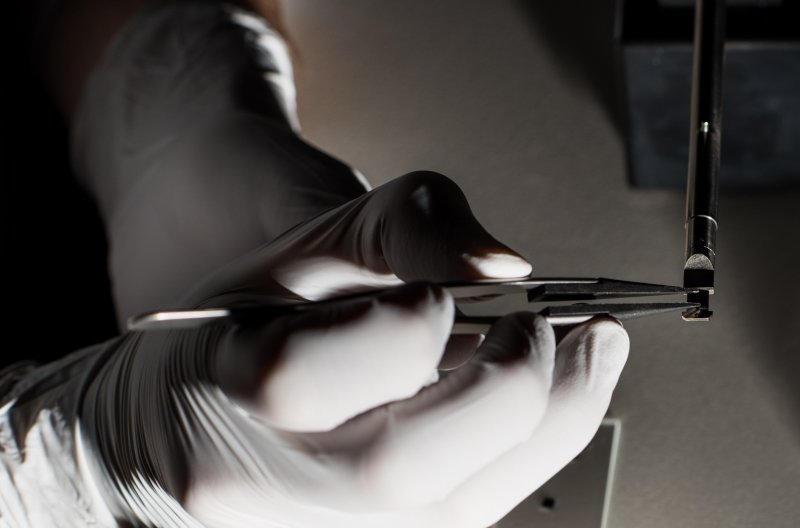
"We have designed and fabricated new devices for holding liquid samples which give us more 'window' regions to collect images than were previously available," Moser says. "Using these multiple windows, we were able to study how the history of electron irradiation influences the nucleation and growth of silver nanoparticles, the growth properties of which are sensitive to the radicals that are generated with the beam. We also used them to study how these radicals impact bacterial cells and demonstrate the extreme sensitivity of these biological samples to the electron beam."
Irradiation from the high-energy beam used in LC-TEM can cause physical damage to samples. For example, the team found that when a cell was imaged—and was exposed to significant electron flux for the first time—observed nanoparticle movement relative to the cell membrane was a result of cell damage. That matters because the insight shows that the movement is an artifact of imaging the cell rather than watching cell dynamics happen in real-time.
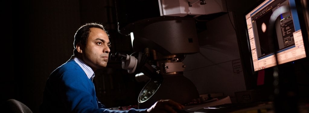
“We were able to capture pristine images of cells using our multi-chamber device wherein the first image represented the cells first exposure to significant electron doses,” Evans says.
"Since the native properties of the sample may be altered or changed by the effects of these electron beam-generated radicals," Shokuhfar adds, "understanding the chemistry changes of a liquid sample as a result of electron irradiation is key to correct interpretation of data collected with this technique."
As the nuances of LC-TEM are gleaned, possible applications include gathering extremely high-resolution, detailed information on energy device and storage materials as well as disease detection, medical imaging and digging deep into the basics of cell activity. In terms of next steps, the team plans to focus on characterizing more biological samples, which appear to be vulnerable to the effects of electron irradiation. The new LC-TEM device offers more windows into this complex atomic world, providing more chances for breakthroughs in energy and health.
Michigan Technological University is an R1 public research university founded in 1885 in Houghton, and is home to nearly 7,500 students from more than 60 countries around the world. Consistently ranked among the best universities in the country for return on investment, Michigan's flagship technological university offers more than 185 undergraduate and graduate degree programs in science and technology, engineering, computing, forestry, business, health professions, humanities, mathematics, social sciences, and the arts. The rural campus is situated just miles from Lake Superior in Michigan's Upper Peninsula, offering year-round opportunities for outdoor adventure.


Comments