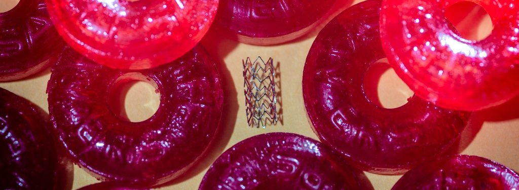

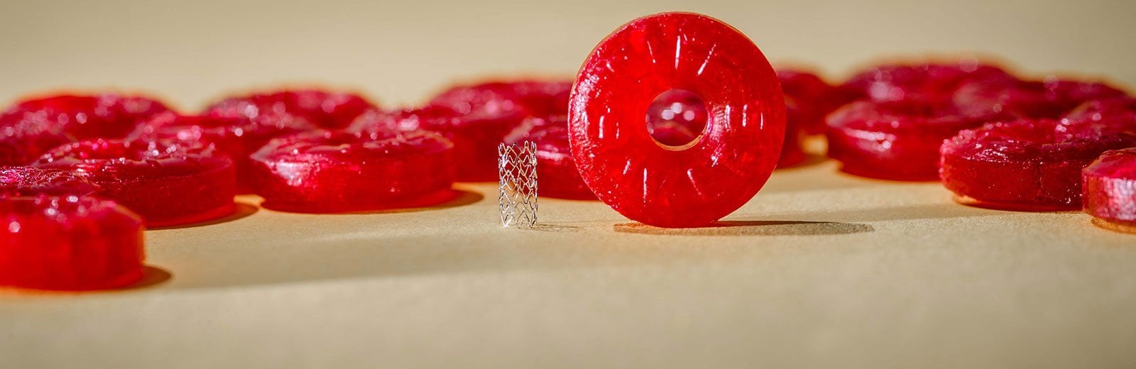
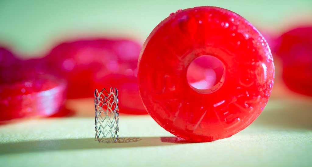
Researchers at Michigan Tech are creating bioabsorbable stents with zinc and zinc alloys that could prove safer than current stent materials.
Building better metallic stents to support weakened arteries has been the work of several years and the focus of a flurry of publications from Michigan Technological University researchers and their collaborators.
The researchers hope to solve a tricky issue in stent construction: The stent itself begins to create more problems than it solves.
Propping Up Arteries Without Damaging Them
Stents are small mesh tubes used to treat narrowed or weakened arteries. Arteries are blood vessels that carry blood away from the heart to other parts of the body. When arteries are blocked or damaged, stents are often used to reopen them. Even though stents can be lifesavers for many, over time the kind of materials the stents are made from can cause other problems to arise, including chronic inflammation, late-stage formation of a blood clot inside a blood vessel, obstruction of blood flow through the circulatory system, and perforation or damage to local cells.
Bodily fluids are extremely corrosive to metallic surfaces; corrosion has traditionally been viewed as one of the major problems for the widely used inert stent metals since the corroded products can cause infections, local pain, swelling, and loosening of the implants. However, advances in biodegradable stent materials whose purpose is to dissolve and be absorbed in the body could address problems related to the permanent presence of traditional stent metals. Manufacturing stents that are biodegradable could revolutionize the way people are treated for clogged arteries, prolonging lives and limiting the need for risky open-heart surgery.
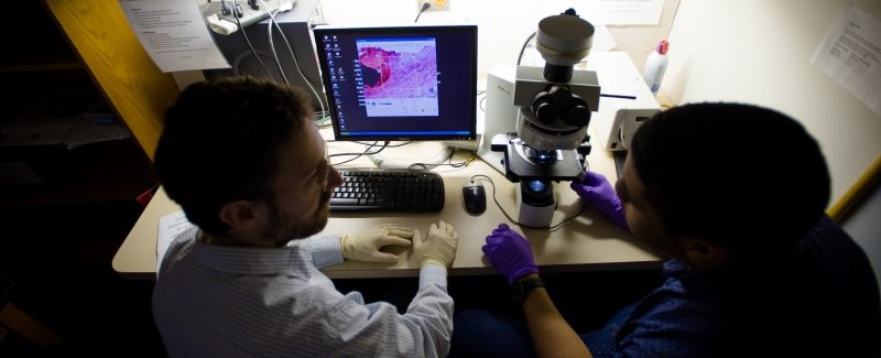
Reaching solutions has been a collaborative effort by scientists in various departments and different fields of expertise, including Jeremy Goldman, Patrick K. Bowen, Jaroslaw Drelich, Sean P. Hopkins, Emily R. Shearier, Elisha J. Earley, Roger J. Guillory, Amani A. Gillette, Eli Aghion, and Martin Bocks, among others. They have relentlessly pushed the boundaries of stent development using zinc.
Designing A Better Stent: An Iterative Process
Driven by the desire to demonstrate the potential for zinc to serve as the base metal for stents and reduce mortality from cardiovascular diseases, the researchers’ first study in 2013 showed that zinc exhibits ideal physiological corrosion behavior for bioabsorbable stents.
Iron and magnesium have been popular stent materials, but the researchers identified—by using a specialized rat-based method—that iron-based metals are prone to rust, and consequently iron was deemed an unsuitable material for coronary stents. For its part, magnesium was found to dissolve too quickly. The unsuitability of iron and magnesium as stent materials called for an alternative metal, hence the shift into metallic zinc studies.
The 2013 discovery of zinc as a breakthrough material heralded the dawn of metallic zinc and its alloys in stent development. With consistent efforts in investigation, the scientists published seven additional impactful papers following up on their work showing zinc’s great promise as a bioabsorbable metal stent compared to all other metals.
The immediate step in the journey was to assess the biological compatibility of the arteries to the metallic zinc to curb the fears of mismatch between cellular tissues and zinc interactions. In 2015, the researchers published that metallic zinc exhibits excellent biocompatibility (which means zinc is not toxic, injurious, or physiologically reactive with living cells to cause immunological rejection) to the arterial cells and tissue. Lack of harmful cellular responses at the stent surface along with tissue regeneration within the biodegradable stent implant as it degraded and became porous, points to the optimal biological acceptance of the zinc.
In 2016, the researchers published numerous papers, each furthering the team’s understanding of the body’s response to stents made of zinc and its related alloys. The first publication in March was sought to assess how zinc affects surrounding cells. Using three different vascular cell types, the researchers published in the ACS Biomaterials and Science Engineering Journal (10.1021/acsbiomaterials.6b00035) that metallic zinc surface adjusted with gelatin (a layer mimicking protein), attached very well with the cells and thrived successfully. Although the initial direct application of the metallic zinc surface (unmodified) to the cells failed to thrive but the attachment itself to the cell was perfect. The cells with zinc modified with the gelatin showed lack of potential toxicity, which was the basis for this study. It was concluded after several techniques used that all the three cell types in this study were able to attach and thrive well on a modified zinc surface, re-assuring biological acceptance and interaction between zinc and the biological bodies. From this study, it was made clearer that the cell attachment, spreading, and movement were affected by increased levels of zinc, particularly in the endothelial and aorta muscle cells.
The researchers published in September 2016 in the journal Surface Innovations (https://doi.org/10.1680/jsuin.16.00014) how different grades and sizes of oxide films were used to manipulate the surface finish of the zinc through different conditions. The results indicated that the rate at which the stent degrades relies more on the stability of the oxide film and its quality. Knowledge gained from this study would guide engineers toward better ways of designing stents and understanding the biocompatibility of zinc oxide as a nanomaterial.
The scientists ended the year 2016 with another publication in ACS Biomaterials Science and Engineering (10.1021/acsbiomaterials.6b00591) on corrosive characteristics that dictate the long-term inflammation of degradable zinc implants. This study examined the inflammatory response of cells to the implants. Using different zinc and zinc alloy materials as implants, the researchers postulated that differences in biocompatibility are due to compositional differences that cause variations. Inflammatory cells could penetrate the thick corrosion layer in the all-zinc implants but the penetration was low in other zinc alloy types with thick enclosure of cells identifying it as foreign material (known as fibrous encapsulation). Hence, the researchers concluded that addition of aluminum to the zinc increases the activity of the localized response (macrophages penetration and growth) thereby generating biologically acceptable inflammatory response. Understanding inflammation boosts the functionality and durability of any implantable device and reduces negative body reactions.
The researchers embraced 2017 by publishing several papers. In the first, the research team investigated zinc and a lithium alloy prepared by melting and hot rolling. They published in Metallurgical and Materials Transactions A (10.1007/s11661-016-3901-0), which demonstrated that the alloy showed similar biocompatibility with cells as seen in the previous study in 2013 using pure zinc, except that the zinc-lithium alloy greatly improved the strength of the stent. Improving stent strength allows it to better support vascular tissues and reduces likelihood of collapse or other related complications.
The second paper was published in the Journal of Biomedical Materials Research Part B (10.1002/jbm.b.33850) on the evaluation of fabricated zinc and an aluminum alloy in stents. The researchers sought a new alloy with better stent application qualities. The researchers observed that the zinc alloy containing up to 5.5 percent aluminum caused inter-granular corrosion (a selective attack near the grain boundaries), which can cause cracking and shattering in implants. Although evaluation showed the zinc-aluminum alloy was biocompatible, the scientists concluded it should be avoided due to the easy cracking.
The final study in 2017 was on the long-term surveillance of a zinc implant in the arteries, which was published in Acta biomaterialia (http://dx.doi.org/10.1016/j.actbio.2017.05.045). The study brought to light that zinc metal implanted in the living artery showed a stable corrosion within 20 months after implantation. This outcome confirmed other predictions that the degradation of zinc stents would require one to two years.
Prior to the discovery of biodegradable stent materials, once a stent is implanted in a patient, it remains for a lifetime, sometimes resulting in the emergence of fatal side effects. Patients with stents implanted in the early 1980s still have them. The scientists from Michigan Tech continue to enhance a biologically acceptable and degradable stent, starting from the discovery of metallic zinc’s applicability to stent construction, working toward improved stent design using zinc alloys.
Michigan Technological University is a public research university founded in 1885 in Houghton, Michigan, and is home to more than 7,000 students from 55 countries around the world. Consistently ranked among the best universities in the country for return on investment, Michigan’s flagship technological university offers more than 120 undergraduate and graduate degree programs in science and technology, engineering, computing, forestry, business and economics, health professions, humanities, mathematics, social sciences, and the arts. The rural campus is situated just miles from Lake Superior in Michigan's Upper Peninsula, offering year-round opportunities for outdoor adventure.

Comments